4 Tests That Detect Esophageal Cancer

April is Esophageal Cancer Awareness Month, which brings to light the roughly 18,500 adults who will receive a diagnosis this year. This cancer that can limit one’s ability to swallow is very lethal, but like with any disease, survival rates improve the earlier it is detected. Here are a few tests doctors can perform during a cancer screening for esophageal cancer.
Standard Diagnostic Tests for Esophageal Cancer
1. Endoscopy & Biopsy
Upper endoscopies are a common method of diagnosing esophageal cancer due to their close-up imaging abilities. For this cancer screening, a patient is given a sedative before the test begins. A doctor then inserts a lighted, flexible tube containing a camera in through the mouth and down the esophagus. The camera then captures a visual of the esophageal lining, noting any abnormalities. If they find anything unusual, they perform a biopsy of the site for further testing.
Lab testing of a biopsy can tell not only if an abnormality is cancer or not, but it also can indicate how far the disease has progressed. Pathologists will study the sample under a microscope, diagnose the type of cancer it is, and assign it a tumor grade to identify how aggressive it is.
2. CT Scan
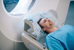 A computed tomography scan, or a CT or CAT scan, uses x-rays to compile complex, cross-sectional images of the body. This testing is essential for tracking if the cancer has spread from the esophagus to other parts of the body. The spread of the disease would likely start at nearby organs or lymph nodes. Doctors will give a patient one to two pints of oral contrast to drink before a CT scan. This liquid helps to emphasize their esophagus and intestines in the imaging to improve the effectiveness of the cancer screening.
3. MRI
Similar to a CT scan, magnetic resonance imaging scans, or MRIs, create detailed imaging of soft tissue. Instead of x-rays, though, this scan uses radio waves and magnets to create the image. MRI scans typically occur to track any abnormalities in the brain and spinal cord, possibly caused by the cancer spreading. A doctor may inject gadolinium, a contrast material, into a vein before the scan to highlight details in the image better.
4. PET Scan
A positron emission tomography scan, or PET scan, typically acts as an auxiliary diagnostic tool during a cancer screening alongside MRI or CT scans. A form of radioactive sugar called fluorodeoxyglucose (FDG) is injected into the blood to identify areas containing cancer cells. FDG isolates these areas because cancer cells tend to absorb more substantial amounts of radioactive sugar than normal cells due to their rapid growth. The radioactivity then shows up on a special camera to track cancer that a doctor believes has spread but can’t identify where. Doctors can sometimes perform PET and CT scans on the same machine, meaning that they can identify areas of higher radioactivity on the PET scan while having the detailed imaging of the CT scan.
When dealing with a potentially critical diagnosis, you want the reassurance that your diagnostics are as accurate as possible. In Flushing, NY, patients rely on Main Street Radiology for excellence in cancer screenings. Accredited by the American College of Radiology, this team of experts will leave no stone unturned to guarantee you receive the most accurate diagnosis possible. For more information about their full range of services, give them a call at (718) 428-1500 or visit their website today.
About the Business

